CurveBeam AI develops advanced musculoskeletal imaging systems for orthopedic specialists. Our portfolio includes industry-leading Weight Bearing CT systems designed to support medical provider diagnostics, treatment planning, and surgical workflows.
CurveBeam AI offers specialized musculoskeletal imaging systems for foot & ankle, knee, hip, distal upper extremity, and surgical planning applications.
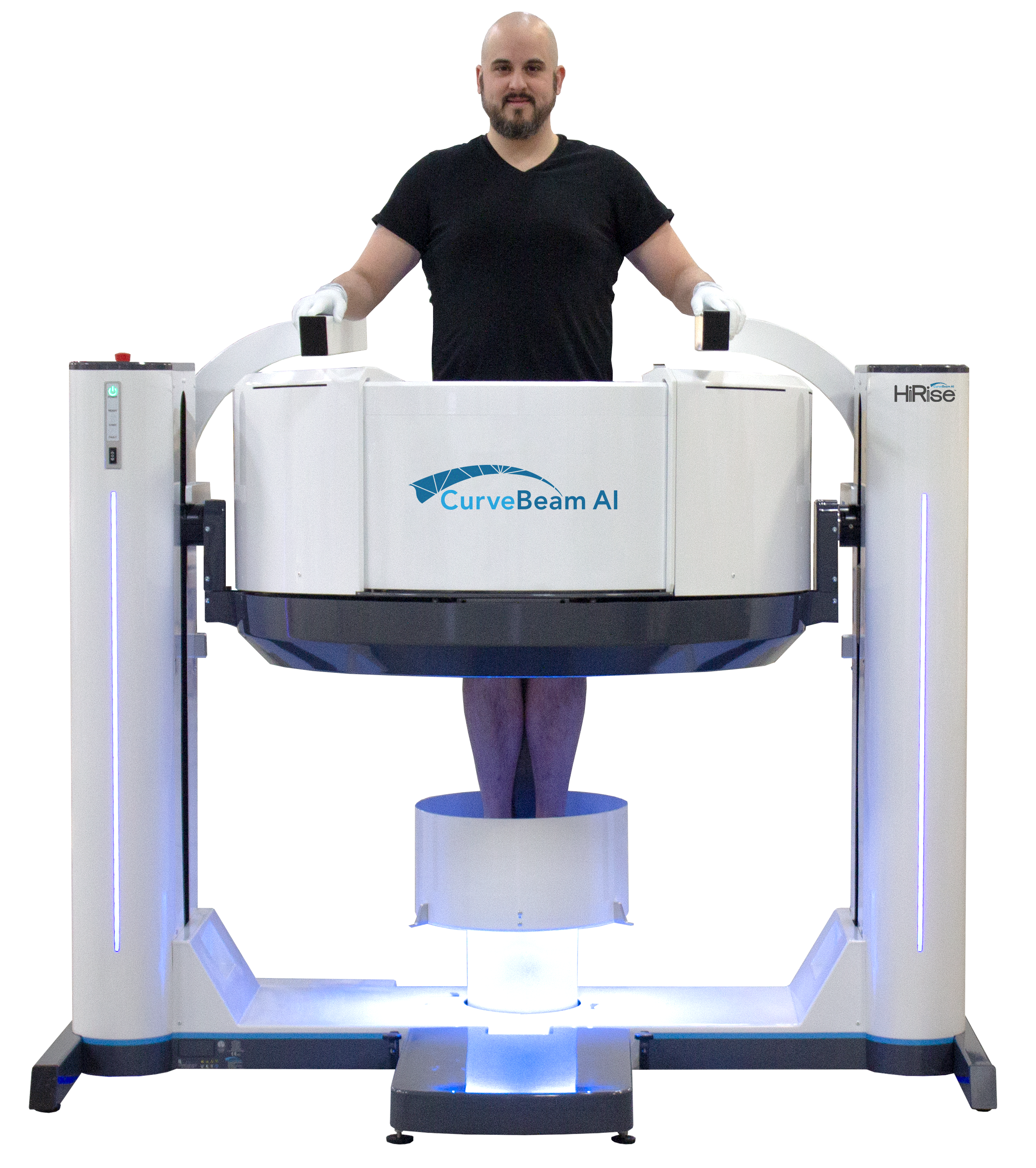
HiRise
Weight bearing CT for standing assessment of the lower extremity
See 3D anatomy and alignment under physiologic load, information conventional supine CT cannot provide.
X-RAY
Weight bearing available
2D projection
CONVENTIONAL CT
3D osseous detail
Typically acquired without physiologic load
MRI
Soft-tissue visualization
Typically acquired without functional loading
WEIGHT BEARING CT
3D osseous anatomy
Acquired under physiologic load
MEDICAL PROVIDER AREAS OF INTEREST
Foot & Ankle
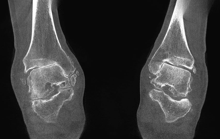
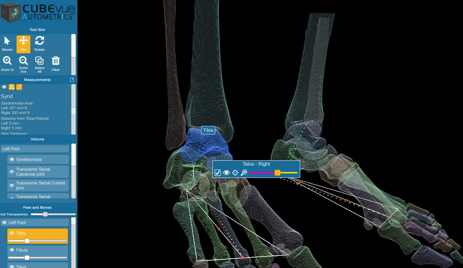
Visualize 3D alignment, joint relationships, and osseous anatomy under physiologic load.
Knee
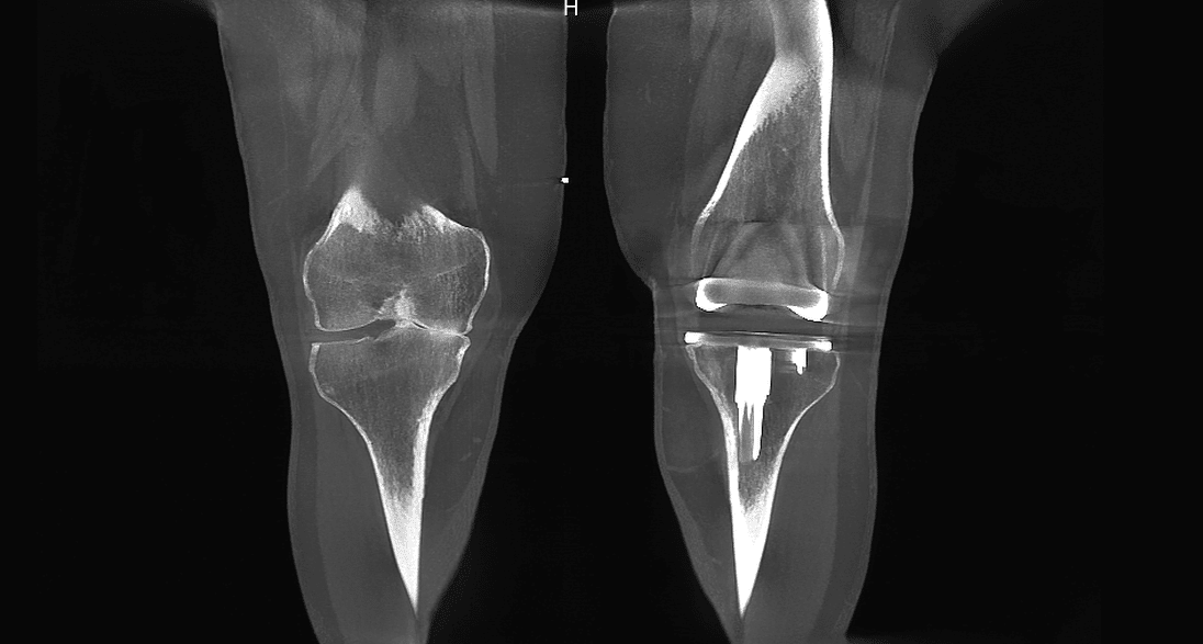
Visualize 3D bony anatomy and lower-extremity alignment in a functional weight-bearing position.
Hip
Visualize hip morphology and alignment with 3D imaging acquired in a functional standing position.
Distal Upper Extremity
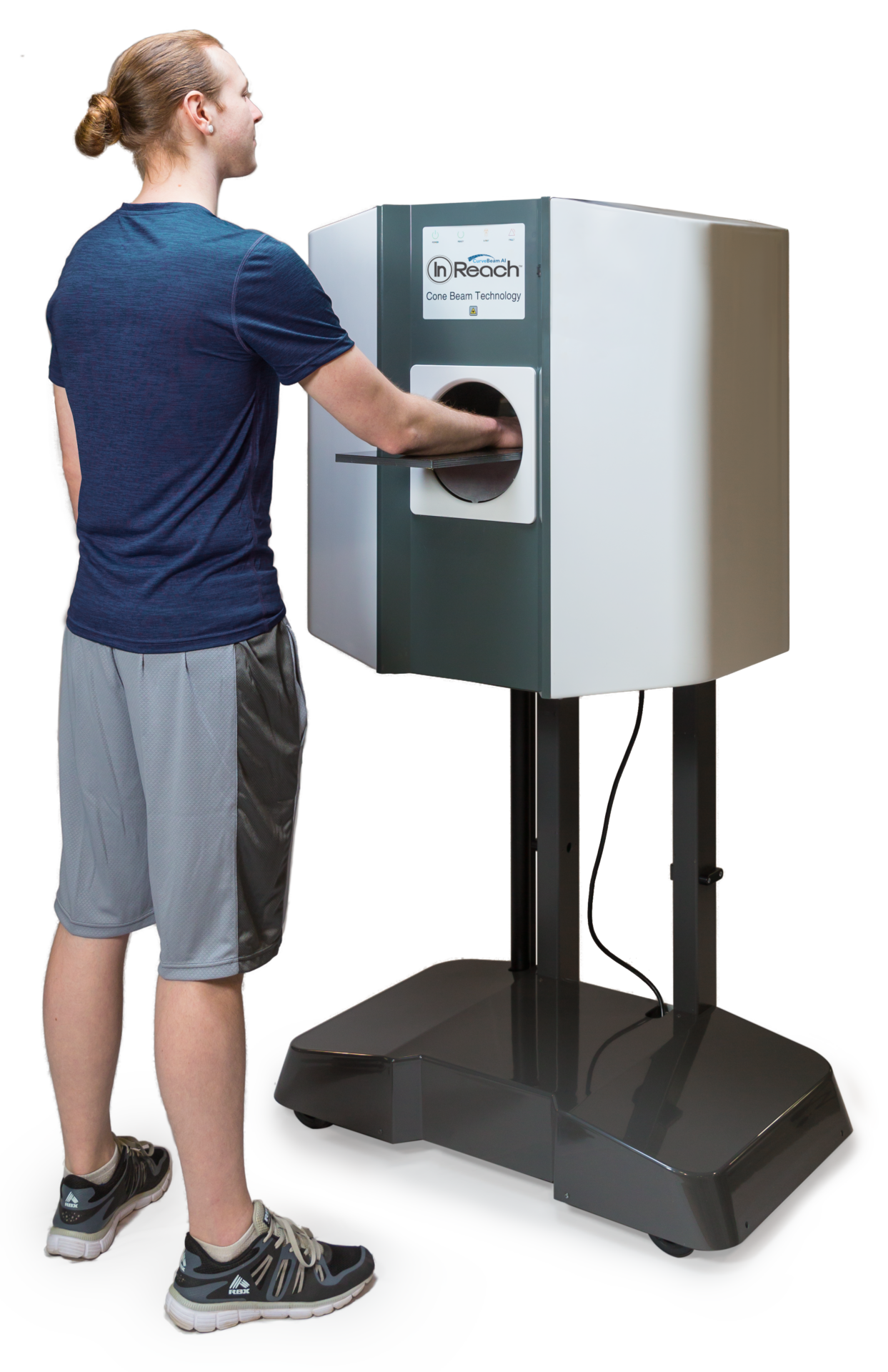
Visualize fractures, alignment, and complex osseous anatomy of the hand, wrist, and elbow in 3D.
Trauma
Visualize fractures and complex osseous anatomy in 3D to support clinical evaluation and treatment planning.
Surgical Planning
Generate CT datasets for supported preoperative planning and patient-specific surgical workflows.
BUILT FOR HOW
MUSCULOSKELETAL
SPECIALISTS PRACTICE
Bring advanced imaging to the point-of-care.
Designed around orthopedic workflows.
Integrate with surgical planning.
Supported Protocols for Vendor Workflows
“We believe every clinical decision deserves the clearest possible picture. Weight-bearing imaging isn’t a luxury. It’s the standard the musculoskeletal field has been waiting for.”
We use cookies on our website to give you the most relevant experience by remembering your preferences and repeat visits. By clicking “Accept All”, you consent to the use of ALL the cookies. However, you may visit "Cookie Settings" to provide a controlled consent.
This website uses cookies to improve your experience while you navigate through the website. Out of these, the cookies that are categorized as necessary are stored on your browser as they are essential for the working of basic functionalities of the website. We also use third-party cookies that help us analyze and understand how you use this website. These cookies will be stored in your browser only with your consent. You also have the option to opt-out of these cookies. But opting out of some of these cookies may affect your browsing experience.
Necessary cookies are absolutely essential for the website to function properly. These cookies ensure basic functionalities and security features of the website, anonymously.
Cookie
Duration
Description
cookielawinfo-checkbox-analytics
11 months
This cookie is set by GDPR Cookie Consent plugin. The cookie is used to store the user consent for the cookies in the category "Analytics".
cookielawinfo-checkbox-functional
11 months
The cookie is set by GDPR cookie consent to record the user consent for the cookies in the category "Functional".
cookielawinfo-checkbox-necessary
11 months
This cookie is set by GDPR Cookie Consent plugin. The cookies is used to store the user consent for the cookies in the category "Necessary".
cookielawinfo-checkbox-others
11 months
This cookie is set by GDPR Cookie Consent plugin. The cookie is used to store the user consent for the cookies in the category "Other.
cookielawinfo-checkbox-performance
11 months
This cookie is set by GDPR Cookie Consent plugin. The cookie is used to store the user consent for the cookies in the category "Performance".
viewed_cookie_policy
11 months
The cookie is set by the GDPR Cookie Consent plugin and is used to store whether or not user has consented to the use of cookies. It does not store any personal data.
Functional cookies help to perform certain functionalities like sharing the content of the website on social media platforms, collect feedbacks, and other third-party features.
Performance cookies are used to understand and analyze the key performance indexes of the website which helps in delivering a better user experience for the visitors.
Analytical cookies are used to understand how visitors interact with the website. These cookies help provide information on metrics the number of visitors, bounce rate, traffic source, etc.
Advertisement cookies are used to provide visitors with relevant ads and marketing campaigns. These cookies track visitors across websites and collect information to provide customized ads.